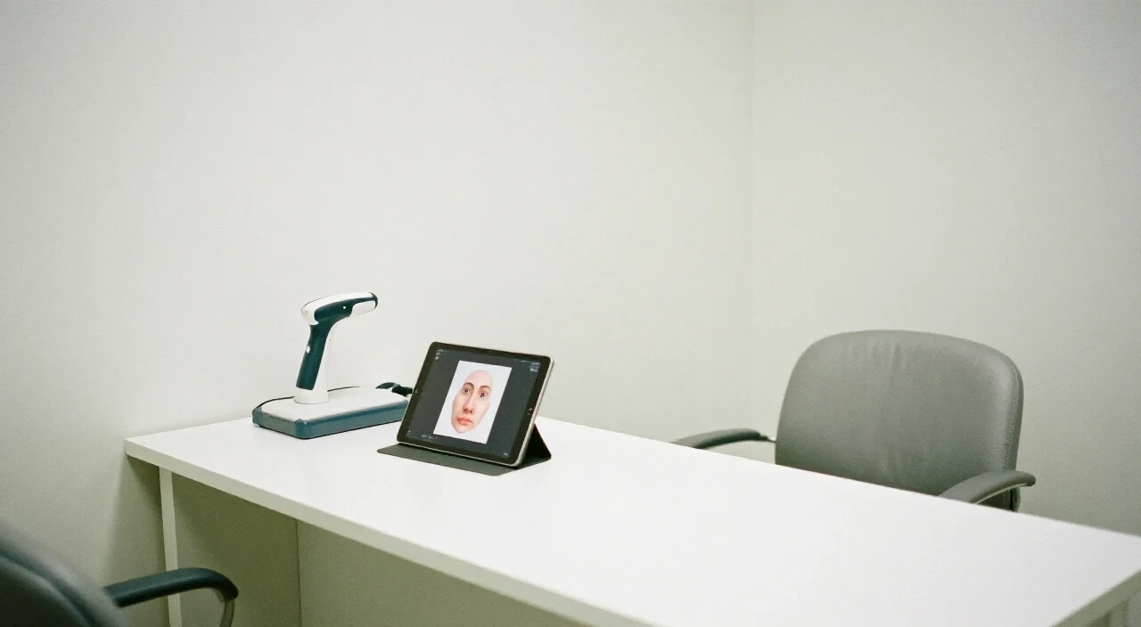
3D imaging rhinoplasty uses digital surface scanning to create a virtual nose simulation that lets patients see possible changes to bridge, tip, profile, projection and alar base before surgery, helping set realistic expectations and improve surgeon-patient communication, though it cannot predict swelling, healing or airflow.
Table of Contents
Key takeaways
- 3D imaging visualises surface changes only
- It improves communication and expectation management
- It cannot model swelling, healing or airway function
- Accurate use requires a physical examination
Is 3D imaging necessary before rhinoplasty?
3D imaging is not mandatory, but it greatly clarifies the aesthetic goals and improves dialogue; most surgeons in Antwerp offer it as part of the initial consultation. While a physical exam remains essential, the visual preview helps patients decide whether to proceed.
The process typically starts with a quick, non-invasive surface scan that captures the nose from multiple angles. The data are processed into a 3-dimensional model, which the surgeon manipulates to show possible adjustments to bridge height, tip rotation, projection and alar base width. Patients can view the changes on a screen or tablet and request minor tweaks in real time.
Limitations include the inability to predict postoperative swelling, scar tissue formation, or how skin thickness will affect the final contour. Therefore, the simulation should be treated as a guide, not a guarantee.
Common mistakes:
- Assuming the displayed image equals the surgical result.
- Choosing a surgeon solely because the software produces attractive pictures.
- Ignoring the need for an internal examination of septum and nasal valves.
When to ask a professional: during the first face-to-face meeting, request a live demonstration of the software and ask how the surgeon integrates the preview with the physical assessment.
Can 3D simulation predict exactly what my nose will look like?
A virtual nose simulation provides an estimate, not a guarantee; it visualizes possible shape changes but cannot account for individual healing, tissue thickness or swelling. Therefore the final appearance may differ, especially in the early postoperative period when swelling is still present.
The algorithm behind the simulation uses the captured surface geometry and applies typical surgical modifications. It does not simulate the biological response of cartilage, bone remodeling, or scar contracture. Consequently, patients with thick skin may see less definition than the image suggests, while those with thin skin may achieve sharper contours.
A 2026 preliminary study found patients rated the influence of 3D simulation at 7.6 out of 10, indicating strong perceived value, yet the same study noted that surgeon communication and before-and-after photographs had an even greater impact on the final decision.
Tips to manage expectations:
- Request to see the simulation side-by-side with pre-operative photos.
- Ask the surgeon how the projected changes translate to tissue movement.
- Remember that the simulation cannot forecast how quickly swelling will subside.
If the preview looks dramatically different from the current anatomy, double-check that the software has correctly aligned the model; misalignment can produce unrealistic results.
Does 3D imaging show breathing problems or internal anatomy?
External 3D imaging captures only surface contours; it does not evaluate the septum, nasal valves, turbinates or airflow, so functional issues must be assessed by physical examination. A surgeon will still perform an internal inspection, possibly with endoscopy, to ensure breathing function is not compromised by the planned aesthetic changes.
The limitation stems from the technology's reliance on light-based scanners that map only the outer skin. Internal structures are hidden behind cartilage and bone, requiring hands-on palpation or imaging such as CT or nasal endoscopy for a complete functional assessment.
During a consultation at Profil Rhinoplasty Atelier, the surgeon combines the virtual simulation with a thorough nasal exam. This includes checking septal deviation, valve competence and turbinate size. If any obstruction is identified, functional correction may be integrated with the aesthetic plan, but it will not be visible in the 3D render.
Common mistake: believing that a beautiful simulated profile guarantees unobstructed breathing. Patients should explicitly ask the surgeon how functional outcomes will be protected.
Checklist for functional evaluation:
- Palpate the nasal bridge for deviation.
- Perform a nasal patency test (e.g., Cottle maneuver).
- Use an endoscope if internal obstruction is suspected.
- Discuss any planned functional procedures alongside the cosmetic changes.
How does 3D imaging affect patient satisfaction and decision-making?
Studies show patients who use 3D simulation report higher satisfaction—about 79% versus 67% without it—because expectations are clearer and decisions are shared with the surgeon. The visual aid also reduces the number of revision surgeries by aligning the surgeon’s plan with the patient’s visual goal from the start.
The 2026 comparative study measured satisfaction three months after surgery and found a statistically significant advantage for the simulation group. Moreover, the same research highlighted that unrealistic or exaggerated images can backfire, leading to disappointment if the real result falls short of the idealised preview.
Profil Rhinoplasty Atelier follows Belgian professional-information rules: the clinic provides factual descriptions of the technology, avoids comparative claims, and does not link the simulation to price promotions. This compliance ensures that patients receive transparent information without misleading advertising.
Effective decision-making steps:
1. Review the virtual simulation together with the surgeon.
2. Discuss which elements are cosmetic versus functional.
3. Identify any aspects of the preview that feel too perfect; ask why.
4. Confirm that the surgeon’s plan includes an internal assessment for breathing.
5. Document the agreed-upon changes in writing before surgery.
When the simulation is used as a communication tool rather than a promise, patients tend to feel more involved and report a smoother postoperative experience.
Practical tip: Treat the virtual nose simulation as a conversation starter, not a contract. Ask the surgeon to explain which parts of the image are based on typical outcomes and which depend on individual healing. This avoids unrealistic expectations and protects against postoperative dissatisfaction.
Frequently asked questions
Is 3D imaging included in the rhinoplasty consultation price?
In Belgium, clinics may list the cost of a consultation, but the inclusion of 3D imaging must be stated as factual information. Many surgeons incorporate the scan at no extra charge, while others list it separately; checking the clinic’s price list clarifies the detail.
Can I compare simulated results from different surgeons?
Yes, but only if each surgeon provides their own scan of the same patient. Because each software may use different rendering algorithms, direct visual comparison can be misleading. Focus on the surgeon’s explanations of how the simulation translates to real tissue changes.
What should I do if the simulation looks too perfect?
Ask the surgeon to point out which features are idealised and which reflect realistic tissue response. An overly flawless preview may indicate that the software has not accounted for skin thickness or postoperative swelling, and it should be tempered with a discussion of likely outcomes.